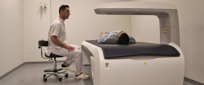
DEXA Scan

Area: Radiology Bone Densitometry or Dual-Energy X-Ray Absorptiometry (DEXA) uses a very small dose of ionizing radiation to produce pictures of the inside of the body (usually the lower spine and hips) to measure bone loss. It is commonly used to diagnose osteoporosis and to assess an individual’s risk for developing fractures. DEXA is Simple Quick

Mammography

Area: Radiology Mammogram is an imaging procedure that is commonly used to detect and diagnose early signs of breast cancer in women. Guidelines for the procedure: DO NOT use the following on your breast and underarms on the day of mammography it is important to avoid them as they will distort the images and will give

Ultrasound

Area: Radiology Ultrasound is a type of imaging. It uses high-frequency sound waves to look at organs and structures inside the body. Health care professionals use it to view the heart, blood vessels, kidneys, liver, and other organs. During pregnancy, doctors use ultrasound to view the fetus. Unlike x-rays, ultrasound does not expose you to radiation.

MRI

Area: Radiology Magnetic Resonance Imaging (MRI) uses a large magnet and radio waves to look at organs and structures inside your body. Health care professionals use MRI scans to diagnose a variety of conditions, from torn ligaments to tumours. MRIs are very useful for examining the brain and spinal cord. Before you get a scan; Your

Fluoroscopy

Area: Radiology Fluoroscopy is a type of medical imaging that shows a continuous X-ray image on a monitor, much like an X-ray movie. During a fluoroscopy procedure, an X-ray beam is passed through the body. The image is transmitted to a monitor so the movement of a body part or of an instrument or contrast agent