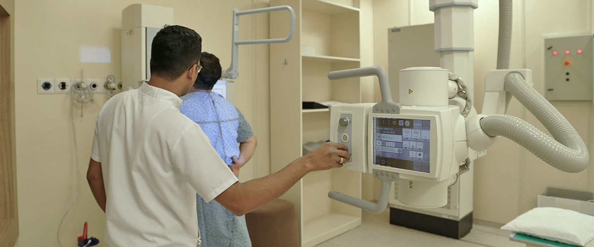
Area: Radiology
X-rays are a type of radiation called electromagnetic waves. This type of imaging creates pictures of the inside of the body. The images show the parts of the body in different shades of black and white. This is because different tissues absorb different amounts of radiation. Calcium in bones absorbs x-rays the most, so bones look white. Fat and other soft tissues absorb less, and look grey. Air absorbs the least, so lungs look black.
The most familiar use of x-rays is checking for broken bones, but x-rays are also used in other ways. For example:
- Chest x-rays can spot pneumonia.
- Mammograms use x-rays to look for breast cancer.
Women should always inform their physician and x-ray technician if there is any possibility that they are pregnant. An x-ray is usually not performed on pregnant women so as not to expose the baby to radiation.
The technician will position the patient on the x-ray table and she/he may be asked to wear a lead shield to help protect certain parts of your body. The x-ray machine will be positioned over the body.
Patient must hold very still and may be asked to keep from breathing for a few seconds while the x-ray picture is taken to reduce the possibility of a blurred image. The technician will walk behind a wall or into the next room to activate the x-ray machine.
The entire x-ray examination, from positioning to obtaining and verifying the images, is usually completed within 15 minutes, although the actual exposure to radiation is usually less than a second.

